The Human Anatomy Laboratory Manual by Elaine Marieb offers a comprehensive guide for understanding human anatomy through hands-on cat dissection exercises. Designed for students, it provides detailed instructions, full-color visuals, and interactive tools to enhance learning. With a focus on practical application, the manual covers all body systems, making complex anatomy accessible and engaging for learners.
1.1 Overview of the Manual
The Human Anatomy Laboratory Manual by Elaine Marieb is a structured guide designed to facilitate a deep understanding of human anatomy through practical dissection exercises. Organized into clear sections, the manual covers all major body systems, with exercises ranging from introductory anatomy to advanced dissections. It incorporates full-color illustrations, photographs, and interactive tools to enhance visual learning. The manual also includes pre-lab activities and safety protocols, ensuring students are well-prepared for hands-on sessions. With its engaging writing style and comprehensive coverage, the manual serves as an essential resource for anatomy students, bridging theory with practical application effectively.
1.2 Importance of Cat Dissections in Anatomy Education
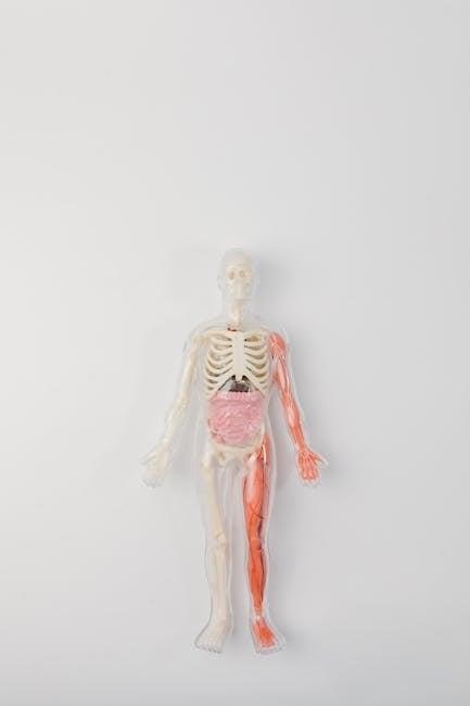
Cat dissections play a pivotal role in anatomy education by providing students with hands-on experience to explore complex anatomical structures. Feline anatomy closely resembles human anatomy, making it an ideal model for studying body systems. Through dissection, students gain a three-dimensional understanding of organ relationships and spatial orientations. This practical approach enhances retention and bridges theoretical knowledge with real-world application. The Marieb Manual emphasizes cat dissections as a safe and ethical way to master anatomical concepts, preparing learners for advanced studies and professional careers in healthcare and biomedical sciences. It ensures a comprehensive and immersive learning experience.
1.3 Key Features of the Marieb Manual
The Marieb Manual is renowned for its comprehensive approach to anatomy education. It features 30 detailed exercises covering all body systems, with full-color illustrations and photographs to enhance visual learning. The manual includes dissection guides, pre-lab and post-lab activities, and interactive digital resources like videos and 3D models. Its clear, engaging writing style makes complex concepts accessible. Additionally, Mastering A&P integration provides online tools for reinforced learning; The combination of hands-on exercises, visual aids, and digital support ensures a well-rounded educational experience, making it an indispensable resource for anatomy students and educators alike.
editions and Updates
The Marieb Manual has evolved through multiple editions, each enhancing content and features. From the 7th to the 9th edition, updates include improved visuals, new exercises, and digital tools, ensuring relevance and effectiveness in anatomy education.
2.1 Overview of the 7th Edition
The 7th edition of the Human Anatomy Laboratory Manual by Elaine Marieb is a comprehensive resource for anatomy students. It features 30 exercises covering all body systems, with a focus on cat dissection. The manual includes full-color illustrations, photographs, and a clear, engaging writing style to facilitate learning. Additional resources such as a companion website and digital tools like pre-lab videos and dissection tutorials enhance student understanding. Designed to support visual and kinesthetic learners, this edition emphasizes hands-on practice and interactive elements, making it an essential tool for anatomy education.
2.2 What’s New in the 8th Edition?
The 8th edition of the Human Anatomy Laboratory Manual by Elaine Marieb introduces several enhancements. New exercises focus on the nervous and digestive systems, providing deeper insights into complex anatomical structures. The manual now includes improved full-color illustrations and interactive 3D models, aiding visual learners. Expanded digital resources, such as dissection videos and pre-lab coaching activities, are accessible via the companion website. Additionally, the 8th edition integrates mobile-friendly tools, enabling students to study on the go. These updates reflect Elaine Marieb’s commitment to advancing anatomy education through innovative and accessible learning materials.
2.3 The 9th Edition: Enhanced Features
The 9th edition of Elaine Marieb’s Human Anatomy Laboratory Manual introduces advanced features to enrich learning. It includes new exercises on the urinary and endocrine systems, offering a more comprehensive understanding. The manual now incorporates enhanced 3D interactive models and expanded dissection videos, providing students with immersive visual aids. Additionally, the 9th edition offers improved organization of content, making it easier to navigate. Elaine Marieb’s commitment to education is evident in these updates, ensuring the manual remains a leading resource for anatomy students. These enhancements reflect her dedication to fostering a deeper and more engaging learning experience.

Pre-Lab Preparation
Prepare for lab by reviewing anatomical terms and procedural steps. Ensure all tools and materials are organized, and maintain a clean, safe workspace for efficient dissection practices.
3.1 Essential Materials and Tools
The manual outlines necessary tools for cat dissection, including scalpels, forceps, and dissecting trays. Essential materials like gloves, preservatives, and anatomical charts are also highlighted. Safety gear such as goggles and lab coats are emphasized to ensure a protected environment. Additionally, the manual provides access to digital resources, including full-color illustrations and interactive models, to enhance understanding. These resources complement hands-on activities, offering a comprehensive learning experience. Proper organization of materials ensures efficient workflow during dissections, making the study of anatomy both effective and engaging for students.
3.2 Safety Protocols and Precautions
Safety is paramount in the anatomy lab. Students must wear personal protective equipment (PPE), including gloves, goggles, and lab coats, to minimize exposure to chemicals and biological specimens. Proper handling and storage of sharp instruments, such as scalpels and forceps, are emphasized to prevent accidents. The manual stresses the importance of working in well-ventilated areas to avoid inhaling preservative fumes. Disposal of biological waste and contaminated materials must follow institutional guidelines to ensure environmental safety. Allergic reactions to preservatives, like formaldehyde, are addressed, urging students to report sensitivities. Adherence to these protocols ensures a safe and structured learning environment for all participants.
3.3 Understanding Anatomical Terminology
Mastering anatomical terminology is essential for effective communication in the lab. Terms like dorsal, ventral, proximal, and distal help locate structures. The manual emphasizes understanding directional terms and body planes to accurately describe dissections. Correct terminology ensures precise identification of organs and tissues, fostering clear communication. This foundation is critical for interpreting instructions and engaging in discussions. By familiarizing themselves with anatomical language, students enhance their learning experience, enabling them to focus on complex dissection procedures with confidence. Proper use of terminology also aids in correlating cat anatomy with human structures, a key feature of the Marieb manual.
Exercises and Body Systems
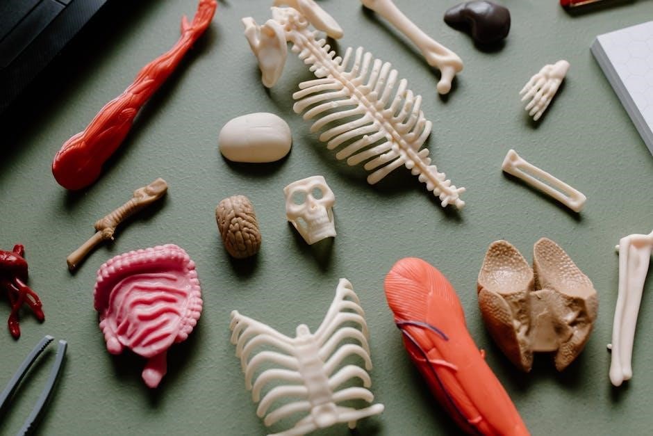
The manual features 30 exercises covering all body systems, with detailed cat dissection guides. Each exercise includes full-color visuals and hands-on activities to enhance understanding of human anatomy. Topics range from the skeletal and muscular systems to the nervous and circulatory systems, providing a comprehensive learning experience that bridges theory and practice effectively.
Exercise 1 provides an overview of laboratory anatomy, emphasizing the importance of proper specimen handling, tool usage, and safety protocols. Students learn to identify key anatomical structures and terminology, laying a foundation for future dissections. The exercise introduces essential lab techniques, such as observation, measurement, and documentation, while fostering a respect for the specimens used in learning. Full-color illustrations and interactive tools support this introduction, ensuring a smooth transition into hands-on anatomy exploration.
4.2 Exercise 2: The Skeletal System
Exercise 2 focuses on the skeletal system, guiding students through the identification and study of bones and their articulations. Using cat specimens, learners explore the structure and function of the axial and appendicular skeleton. Full-color illustrations and dissection videos enhance understanding, while interactive 3D models allow for detailed examination of bone anatomy. Activities include labeling bones, observing joints, and comparing feline and human skeletal systems. This exercise provides a foundational understanding of skeletal anatomy, preparing students for more complex dissections and body system studies in subsequent exercises. Hands-on exploration fosters a deeper appreciation of the skeletal system’s role in movement and support.
4.3 Exercise 3: The Muscular System
Exercise 3 delves into the muscular system, focusing on the identification and exploration of major muscle groups in cat specimens. Students learn to dissect and examine feline musculature, comparing it to human anatomy. The manual provides detailed illustrations and dissection videos to aid in understanding muscle structure, origin, and insertion points. Activities include identifying muscles of movement and observing their functions. Interactive 3D models further enhance the study of muscle groups and their roles in movement. This exercise helps students appreciate the complexity of the muscular system and its essential role in body movement and stability, bridging feline and human anatomical comparisons.
4.4 Exercise 4: The Nervous System
Exercise 4 focuses on the nervous system, guiding students through the dissection of a cat’s nervous structures to understand their organization and function. The manual includes detailed step-by-step instructions for identifying key components such as the brain, spinal cord, and peripheral nerves. High-quality illustrations and dissection videos complement the lab work, helping students visualize nerve pathways and connections. Interactive 3D models allow exploration of neural structures, enhancing comprehension of how the nervous system integrates and controls body functions. This exercise bridges feline anatomy with human nervous system principles, providing a practical foundation for advanced study in neuroscience and related fields.
4.5 Exercise 5: The Circulatory System
Exercise 5 delves into the circulatory system, providing a detailed dissection guide for examining the cat’s heart, blood vessels, and lymphatic structures. Students learn to identify the atria, ventricles, and major arteries, correlating feline anatomy with human physiology. The manual includes full-color illustrations and dissection videos to aid in visualizing complex vascular networks. Interactive 3D models further enhance understanding of blood flow and circulation. This exercise emphasizes the functional relationships between the heart, lungs, and peripheral vessels, equipping learners with practical skills in anatomical identification and reinforcing key concepts in cardiovascular anatomy through hands-on exploration.
4.6 Exercise 6: The Respiratory System
Exercise 6 focuses on the respiratory system, guiding students through the dissection of a cat’s nasal passages, trachea, bronchi, and lungs. The manual provides detailed instructions for identifying key structures, such as the epiglottis, larynx, and diaphragm. Full-color illustrations and dissection videos enhance visualization, while interactive 3D models demonstrate airflow and gas exchange. This exercise emphasizes the functional relationship between the respiratory and circulatory systems, allowing learners to explore how oxygenation occurs. Practical skills in identifying respiratory anatomy are reinforced, making complex concepts accessible and engaging for anatomy students.
4.7 Exercise 7: The Digestive System
Exercise 7 delves into the digestive system, focusing on the anatomy of a cat’s gastrointestinal tract. Students explore the oral cavity, esophagus, stomach, small intestine, and large intestine. The manual provides detailed dissection instructions, accompanied by full-color illustrations and dissection videos. Key structures like the liver, pancreas, and gallbladder are examined, highlighting their roles in digestion. Interactive 3D models further clarify the spatial relationships between organs. This exercise emphasizes the functional connections between digestive structures, enabling students to understand nutrient absorption and waste elimination. Practical dissection skills are reinforced, making the digestive system’s complexity more accessible and engaging for learners.
4.8 Exercise 8: The Urinary System
Exercise 8 explores the urinary system through cat dissection, focusing on the kidneys, ureters, bladder, and urethra. Students examine the renal corpuscle, nephrons, and urinary tract structures. Full-color illustrations and dissection videos guide learners through identifying these components. The exercise emphasizes the relationship between the urinary and reproductive systems, as well as the role of the kidneys in filtration and waste elimination. Interactive 3D models further aid in understanding the spatial arrangement of urinary organs. This hands-on approach helps students grasp the functional anatomy of the urinary system, preparing them for clinical applications and advanced studies in anatomy.
4.9 Exercise 9: The Reproductive System
Exercise 9 delves into the reproductive system, examining the male and female cat anatomy. Students identify structures such as the ovaries, uterus, testes, and accessory glands. Full-color illustrations and videos aid in understanding reproductive organs and their functions. The exercise highlights the interface between the urinary and reproductive systems, emphasizing anatomical differences and physiological roles. Interactive 3D models and pre-lab videos enhance comprehension, allowing learners to visualize complex structures. This practical exploration provides a foundation for understanding human reproductive anatomy, essential for future clinical studies and healthcare professions. The manual’s engaging approach ensures a comprehensive learning experience.
4.10 Exercise 10: The Endocrine System
Exercise 10 focuses on the endocrine system, exploring glands and their hormonal functions. Students examine the pancreas, thyroid, adrenal glands, and others, linking anatomy to physiology. Full-color images and dissection videos clarify glandular structures and their roles. Interactive 3D models enhance understanding of hormone production and regulation. The exercise emphasizes the endocrine system’s integration with other body systems, such as the nervous and reproductive systems. Pre-lab videos and activities in Mastering A&P provide additional support, ensuring a thorough grasp of endocrine anatomy and its significance in human health and disease. This exercise bridges theoretical knowledge with practical observation.
Cat Dissection Process
The cat dissection process provides a hands-on learning experience, guiding students through systematic exploration of anatomy. Essential tools and safety protocols ensure a safe, educational experience, aligning with manual content and laboratory goals.
5.1 Step-by-Step Guide to Cat Dissection
The Marieb manual provides a detailed, step-by-step guide for cat dissection, ensuring a systematic approach to exploring anatomy. Each exercise begins with an external examination, followed by skin removal and identification of superficial structures. Students then progress to deeper dissections, carefully exposing organs and systems. Essential tools like scalpels, forceps, and dissecting trays are emphasized. The guide incorporates visual aids, such as full-color illustrations and dissection videos, to enhance understanding. Safety protocols and proper specimen handling are highlighted throughout. This hands-on process fosters a practical understanding of anatomical relationships and prepares students for advanced studies in human anatomy and physiology.
5.2 Preserving Specimens for Future Use
Preserving cat specimens for future use involves proper techniques to maintain anatomical integrity. The manual recommends using formalin for fixation, ensuring specimens remain intact and suitable for repeated dissections. Storage in sealed containers with preservative solution prevents decay. Proper labeling and organization are emphasized for easy access. Safety guidelines, such as wearing gloves and working in ventilated areas, are highlighted. Disposal must follow biohazard protocols to ensure environmental and health safety. These steps ensure specimens remain usable for extended periods, supporting ongoing educational and research needs in anatomy labs. The manual provides detailed instructions to maximize specimen longevity and safety.
5.3 Common Challenges and Solutions
During cat dissections, students often face challenges such as specimen degradation and difficulty identifying structures. Proper preservation techniques, like formalin fixation, help maintain specimen quality. Using magnification tools and reference images can enhance structure identification. Safety concerns, like exposure to chemicals, are mitigated with protective gear and ventilation. Dissection guides and instructor support also aid in overcoming technical difficulties. Engaging with supplementary resources, such as videos and 3D models, can improve understanding and address common challenges effectively, ensuring a successful and safe learning experience in anatomy labs.
Visual Aids and Resources
The manual includes full-color illustrations, photographs, and dissection videos to enhance understanding. Interactive 3D models and Mastering A&P provide additional tools for engaging and effective anatomy learning.
6.1 Full-Color Illustrations and Photographs
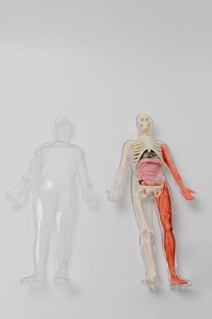
The Human Anatomy Laboratory Manual features full-color illustrations and photographs that provide detailed visual representations of anatomical structures. These visuals aid students in understanding complex dissections and comparing human anatomy with cat specimens. The images are carefully labeled to highlight key features, making them invaluable for hands-on learning. The manual’s clear and engaging writing style is complemented by these high-quality visuals, ensuring a comprehensive understanding of each exercise. The illustrations are particularly effective in showcasing the similarities and differences between human and feline anatomy, making them an essential resource for both classroom and laboratory settings. This visual approach enhances the learning experience, fostering deeper anatomical knowledge.
6.2 Dissection Videos and Tutorials
The manual is supported by dissection videos and tutorials that provide step-by-step guidance for students. These resources include pre-lab coaching activities, detailed dissection demonstrations, and interactive tutorials. The videos cover key exercises, such as cat dissections, offering a visual learning experience that complements the written content. They are particularly useful for understanding complex anatomical structures and procedures. By combining visual and hands-on learning, these tutorials enhance students’ mastery of human anatomy. The videos are accessible online, making them a flexible and valuable tool for both in-lab and at-home study, ensuring a comprehensive understanding of each exercise. This feature enriches the learning process significantly.
6.3 Interactive 3D Models and Tools
The Human Anatomy Laboratory Manual features interactive 3D models and tools that enhance the learning experience. These digital resources allow students to explore anatomical structures in detail, with the ability to rotate, zoom, and label parts of 3D models. The tools are particularly useful for visualizing complex systems and their relationships. They complement traditional dissection exercises by providing a virtual environment for practice and review. Accessible online, these interactive models are a valuable supplement to the manual, offering a modern and engaging way to study anatomy. They make learning more interactive and effective for students of all skill levels. These tools are integral to the manual’s comprehensive approach to anatomy education.
Author Background
Elaine Nicpon Marieb, a renowned anatomy educator, is celebrated for her clear, engaging teaching style. Her work emphasizes practical learning, benefiting students worldwide through her laboratory manuals and philanthropic efforts.
7.1 Elaine Marieb: Biography and Contributions
Elaine Nicpon Marieb, a distinguished educator and author, revolutionized anatomy education with her laboratory manuals. Her work integrates clear instructions with visual aids, making complex concepts accessible. Renowned for her Human Anatomy Laboratory Manual with Cat Dissections, Marieb emphasizes hands-on learning through dissection exercises. Her contributions extend beyond academia; the Elaine Nicpon Marieb Charitable Foundation supports educational opportunities for students. Marieb’s dedication to teaching and her innovative approach have left a lasting impact on anatomy education, benefiting students and educators globally.
7.2 The Elaine Nicpon Marieb Charitable Foundation
The Elaine Nicpon Marieb Charitable Foundation, established by Dr. Marieb, focuses on advancing education and supporting students in anatomy and physiology. The foundation provides scholarships, grants, and resources to students pursuing careers in science and healthcare. It also supports educational initiatives to enhance teaching and learning in anatomy. Through her philanthropy, Dr. Marieb has made a significant impact on the scientific community, ensuring access to quality education for future professionals. This reflects her commitment to fostering excellence in anatomy education and her dedication to giving back to the field she has profoundly influenced.
7.3 Dr. Marieb’s Impact on Anatomy Education
Dr. Elaine Marieb has revolutionized anatomy education through her groundbreaking laboratory manuals and textbooks. Her work has transformed how students and educators approach anatomy, making complex concepts accessible and engaging. The inclusion of cat dissections in her manuals has provided a practical, hands-on learning experience, bridging the gap between theoretical knowledge and real-world application. Her clear, concise writing style and the integration of visual aids have set a new standard in anatomy education. Dr. Marieb’s contributions have inspired generations of students and educators, leaving a lasting legacy in the field of anatomy and physiology.

Digital and Supplementary Resources
The manual offers a companion website with online access, Mastering A&P interactive features, dissection videos, and mobile apps, enhancing learning through digital engagement and convenience.
8.1 Companion Website and Online Access
The Companion Website for the Marieb Human Anatomy Laboratory Manual provides a wealth of digital resources to support student learning. Accessible online, it includes pre-lab video coaching, interactive activities, and dissection videos to enhance understanding. Students can explore 3D models, quizzes, and additional study materials tailored to each exercise. The website offers a user-friendly interface, allowing learners to access content anytime, anywhere. It complements the lab manual by providing engaging tools that reinforce key concepts and promote active learning. This digital support ensures students are well-prepared for lab sessions and beyond.
8.2 Mastering A&P: Interactive Features
Mastering A&P enhances the learning experience with interactive tools tailored for the Marieb lab manual. Features include 3D bone and muscle models, virtual dissection simulations, and adaptive quizzes. Video tutorials guide students through complex procedures, while real-time feedback helps track progress. The platform supports active learning, catering to diverse learning styles. Instructors can customize assignments to align with lab exercises, ensuring a cohesive educational experience. This integration of technology and anatomy fosters deeper understanding and engagement, making it an invaluable resource for both students and educators in anatomy education;
8.3 Mobile Apps for On-the-Go Learning
Mobile apps complement the Marieb lab manual, offering flexible learning opportunities. Features include flashcards, interactive quizzes, and 3D anatomical models. Students can access dissection tutorials and review body systems anytime, anywhere. Offline functionality ensures learning continues without internet connectivity. Customizable study plans and progress tracking further enhance engagement. These apps bridge classroom and lab experiences, providing a convenient way to reinforce anatomy concepts. They are designed to support active learning and cater to diverse study preferences, making anatomy education more accessible and effective for modern learners.
The Marieb manual effectively bridges theory and practice, offering a comprehensive, interactive approach to anatomy education. Its engaging tools and clear guidance ensure lasting understanding for learners.
9.1 Summary of Key Benefits
The Human Anatomy Laboratory Manual with cat dissections by Elaine Marieb offers a comprehensive, hands-on approach to anatomy education. With 30 exercises covering all body systems, it provides a clear, engaging writing style and full-color illustrations to enhance understanding. The manual includes dissection videos, interactive 3D models, and pre-lab coaching activities, making complex concepts accessible. Its practical focus on cat dissections allows students to explore anatomical structures in a real-world context. Additionally, the companion website and mobile apps provide supplementary resources for on-the-go learning, ensuring a well-rounded educational experience. This manual is an invaluable tool for anatomy students seeking both depth and clarity in their studies.
9.2 Future Directions in Anatomy Education
Future anatomy education will likely integrate advanced technologies like virtual dissections and augmented reality to enhance learning. The Marieb manual’s digital resources, such as Mastering A&P and 3D models, already pave the way for interactive learning. As educators embrace personalized learning, adaptive assessments and real-time feedback tools will become more prevalent. The Elaine Nicpon Marieb Charitable Foundation supports innovations in anatomy education, ensuring access to cutting-edge resources. With a focus on global accessibility and interactive engagement, anatomy education will continue to evolve, providing students with immersive and effective learning experiences that bridge traditional and digital methods seamlessly.